Sharp Focusing Made Easy – X-ray Lenses with Built-in Error Correction Developed
X-ray lenses reveal tiny details, either by providing complete images or by enabling precise scanning. They are used in PETRA IV research to identify objects that remain invisible to normal microscopes.

X-ray Lenses with Built-in Error Correction Developed
An international team of researchers, including scientists from DESY, has developed a new type of X-ray optics for large-scale research facilities. These optics allow X-rays to be focused down to a spot just 50 nanometers wide—without the need for complex adjustment or alignment. The new lenses represent an important advancement for cutting-edge X-ray analysis, such as what will be possible with PETRA IV.
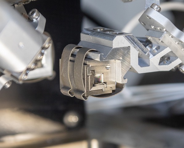
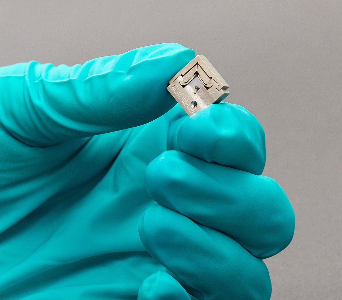
The team created special tiny diamond “lens cubes” that act as lenses for X-rays. These lenses are particularly useful for high-resolution imaging in materials science and biology, where they help researchers examine minute structures—such as inside cells, new materials, or chemical reactions.
The DESY team has now succeeded in manufacturing the lenses with such precision that they focus X-rays optimally without requiring post-installation calibration. Typically, optical systems like these need to be painstakingly aligned after installation to perform at their best. With this new DESY design, that step is no longer necessary.
The high image quality was confirmed using a standard test pattern, demonstrating resolution of structures down to 50 nanometers.
“As far as we know, this is the smallest focus ever achieved with diamond lenses,” says Wenxin Wang, DESY Scientist and lead author of the study.

High-Precision Manufacturing
The diamond lenses were fabricated using highly precise laser techniques, which is key to their performance. They are designed to automatically correct optical errors—known as aberrations—resulting in sharper and more accurate images. Another advantage: the correction mechanisms are built directly into the lenses, eliminating the need for additional correction components.
“We’ve developed a system that can deliver high-resolution X-ray images very quickly, which makes it much easier to use in the lab,” says Frank Seiboth, a scientist at DESY. “It saves time and simplifies working with high-resolution X-ray imaging.”
The new technology was tested at DESY’s PETRA III research light source, specifically at beamline P06, which is equipped with the Ptychographic Nano-Analytical Microscope (PtyNAMi). This facility enables nanometer-scale high-resolution X-ray imaging.
Link to the publication:
Wenxin Wang, Ralph Döhrmann, Stephan Botta, Anders Madsen, Christian G. Schroer, and Frank Seiboth, Diamond X-ray lens cubes with integrated aberration compensation, Opt. Express 33 (2025), DOI: 10.1364/OE.562556

